Abstract
Case Report
Carotid artery disease: AngioCT features
Joana Ferreira*, Olinda Miranda, Alexandre Lima Carneiro, Sandrina Braga, João Correia Simões, Celso Carrilho, Amílcar Mesquita and Jorge Cotter
Published: 26 July, 2019 | Volume 3 - Issue 2 | Pages: 059-060
The objective of this paper is to emphasis the AngioCT features of carotid dissection/mural hematoma. The image show an internal carotid artery with narrowly eccentric lumen surrounded by a crescent-shaped hypodense mural thickening, with a visibly enhancing vessel wall. The carotid hematoma is a hypodense mural thickening that leads to expansion of the arterial wall, compression of the lumen and release of thrombogenic factors by intimal damage. Hematoma between the intima and media causes vessel expansion diameter and a narrow eccentric lumen. Peripheral hyper density is due to the contrast enhancement of the vasa vasorum in the adventitial layer. The physician should be familiar with the imagiologic features of carotid arterial disease, due to the diferent treatment options.
Read Full Article HTML DOI: 10.29328/journal.ascr.1001035 Cite this Article Read Full Article PDF
Keywords:
Carotid artery; Dissection; Mural hematoma; Atherosclerosis; AngioCT; Stenosis
References
- Patel RR, Adam R, Maldjian C, Lincoln CM, Yuen A, et al. Cervical Carotid Artery Dissection: Current Review of Diagnosis and Treatment. Cardiol Rev. 2012; 20: 145-152. PubMed: https://www.ncbi.nlm.nih.gov/pubmed/22301716
- Rodallec MH, Marteau V, Gerber S, Desmottes L, Zins M. Craniocervical Arterial Dissection: Spectrum of Imaging Findings and Differential Diagnosis. Radiographics. 2008; 28: 1711-1728. PubMed: https://www.ncbi.nlm.nih.gov/pubmed/18936031
- Vilela P, Goulão A. Cervical and intracranial arterial dissection: review of the acute clinical presentation and imaging of 48 cases. Acta Med Port. 2003; 16: 155-164. PubMed: https://www.ncbi.nlm.nih.gov/pubmed/12868394
- Thanvi B, Munshi SK, Dawson SL, Robinson TG. Carotid and Vertebral Artery Dissection Syndromes. Postgrad Med J. 2005; 81: 383-388. PubMed: https://www.ncbi.nlm.nih.gov/pubmed/15937204
- Selwaness M, Bouwhuijsen Q, Onkelen R, Hofman A, Franco O, et al. Atherosclerotic Plaque in the Left Carotid Artery Is More Vulnerable Than in the Right. Stroke. 2014; 45: 3226-3230. PubMed: https://www.ncbi.nlm.nih.gov/pubmed/25228259
- U-King-Im JM, Fox AJ, Aviv RI, Howard P, Yeung R, et al. Characterization of Carotid Plaque Hemorrhage. a CT Angiography and MR Intraplaque Hemorrhage Study. Stroke. 2010; 41: 1623-1629. PubMed: https://www.ncbi.nlm.nih.gov/pubmed/20576955
Figures:
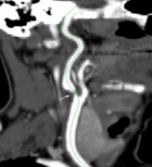
Figure 1
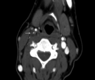
Figure 2
Similar Articles
-
Gossypiboma due to a retained surgical sponge following abdominal hysterectomy, complicated by intestinal migration and small bowel obstruction- A Case ReportVivek Agrawal,Praroop Gupta*. Gossypiboma due to a retained surgical sponge following abdominal hysterectomy, complicated by intestinal migration and small bowel obstruction- A Case Report. . 2018 doi: 10.29328/journal.ascr.1001017; 2: 015-017
-
Laparoscopic Cholecystectomy: Challenges faced by beginners our perspectiveKunal Chowdhary,Gurinder Kaur,Kapil Sindhu,Muzzafar Zaman*,Aliya Shah,Rohit Dang,Ashish Kumar,Jose John Maiakal,Ashutosh Bawa. Laparoscopic Cholecystectomy: Challenges faced by beginners our perspective. . 2018 doi: 10.29328/journal.ascr.1001018; 2: 018-024
-
Carotid artery disease: AngioCT featuresJoana Ferreira*,Olinda Miranda,Alexandre Lima Carneiro,Sandrina Braga,João Correia Simões,Celso Carrilho, Amílcar Mesquita,Jorge Cotter. Carotid artery disease: AngioCT features. . 2019 doi: 10.29328/journal.ascr.1001035; 3: 059-060
-
Beginnings of bariatric and metabolic surgery in SpainAniceto Baltasar*. Beginnings of bariatric and metabolic surgery in Spain. . 2019 doi: 10.29328/journal.ascr.1001042; 3: 082-090
-
Stomach cancer: epidemiological, diagnostic and therapeutic aspects at the Kara Teaching Hospital, TogoDOSSOUVI Tamegnon*,EL-HADJI YAKOUBOU Rafiou,ADABRA Komlan,AMAVI Ayi,AMOUZOU Efoé-Ga Olivier,KANASSOUA Kokou Kouliwa,KASSEGNE Iroukora,DOSSEH Ekoué David. Stomach cancer: epidemiological, diagnostic and therapeutic aspects at the Kara Teaching Hospital, Togo. . 2022 doi: 10.29328/journal.ascr.1001062; 6: 001-003
-
CVS: An Effective Strategy to Prevent Bile Duct InjurySardar Rezaul Islam*, Debabrata Paul, Shah Alam Sarkar, Mohammad Hanif Emon, Tania Ahmed. CVS: An Effective Strategy to Prevent Bile Duct Injury. . 2024 doi: 10.29328/journal.ascr.1001080; 8: 027-031
Recently Viewed
-
Agriculture High-Quality Development and NutritionZhongsheng Guo*. Agriculture High-Quality Development and Nutrition. Arch Food Nutr Sci. 2024: doi: 10.29328/journal.afns.1001060; 8: 038-040
-
A Low-cost High-throughput Targeted Sequencing for the Accurate Detection of Respiratory Tract PathogenChangyan Ju, Chengbosen Zhou, Zhezhi Deng, Jingwei Gao, Weizhao Jiang, Hanbing Zeng, Haiwei Huang, Yongxiang Duan, David X Deng*. A Low-cost High-throughput Targeted Sequencing for the Accurate Detection of Respiratory Tract Pathogen. Int J Clin Virol. 2024: doi: 10.29328/journal.ijcv.1001056; 8: 001-007
-
A Comparative Study of Metoprolol and Amlodipine on Mortality, Disability and Complication in Acute StrokeJayantee Kalita*,Dhiraj Kumar,Nagendra B Gutti,Sandeep K Gupta,Anadi Mishra,Vivek Singh. A Comparative Study of Metoprolol and Amlodipine on Mortality, Disability and Complication in Acute Stroke. J Neurosci Neurol Disord. 2025: doi: 10.29328/journal.jnnd.1001108; 9: 039-045
-
Development of qualitative GC MS method for simultaneous identification of PM-CCM a modified illicit drugs preparation and its modern-day application in drug-facilitated crimesBhagat Singh*,Satish R Nailkar,Chetansen A Bhadkambekar,Suneel Prajapati,Sukhminder Kaur. Development of qualitative GC MS method for simultaneous identification of PM-CCM a modified illicit drugs preparation and its modern-day application in drug-facilitated crimes. J Forensic Sci Res. 2023: doi: 10.29328/journal.jfsr.1001043; 7: 004-010
-
A Gateway to Metal Resistance: Bacterial Response to Heavy Metal Toxicity in the Biological EnvironmentLoai Aljerf*,Nuha AlMasri. A Gateway to Metal Resistance: Bacterial Response to Heavy Metal Toxicity in the Biological Environment. Ann Adv Chem. 2018: doi: 10.29328/journal.aac.1001012; 2: 032-044
Most Viewed
-
Evaluation of Biostimulants Based on Recovered Protein Hydrolysates from Animal By-products as Plant Growth EnhancersH Pérez-Aguilar*, M Lacruz-Asaro, F Arán-Ais. Evaluation of Biostimulants Based on Recovered Protein Hydrolysates from Animal By-products as Plant Growth Enhancers. J Plant Sci Phytopathol. 2023 doi: 10.29328/journal.jpsp.1001104; 7: 042-047
-
Sinonasal Myxoma Extending into the Orbit in a 4-Year Old: A Case PresentationJulian A Purrinos*, Ramzi Younis. Sinonasal Myxoma Extending into the Orbit in a 4-Year Old: A Case Presentation. Arch Case Rep. 2024 doi: 10.29328/journal.acr.1001099; 8: 075-077
-
Feasibility study of magnetic sensing for detecting single-neuron action potentialsDenis Tonini,Kai Wu,Renata Saha,Jian-Ping Wang*. Feasibility study of magnetic sensing for detecting single-neuron action potentials. Ann Biomed Sci Eng. 2022 doi: 10.29328/journal.abse.1001018; 6: 019-029
-
Pediatric Dysgerminoma: Unveiling a Rare Ovarian TumorFaten Limaiem*, Khalil Saffar, Ahmed Halouani. Pediatric Dysgerminoma: Unveiling a Rare Ovarian Tumor. Arch Case Rep. 2024 doi: 10.29328/journal.acr.1001087; 8: 010-013
-
Physical activity can change the physiological and psychological circumstances during COVID-19 pandemic: A narrative reviewKhashayar Maroufi*. Physical activity can change the physiological and psychological circumstances during COVID-19 pandemic: A narrative review. J Sports Med Ther. 2021 doi: 10.29328/journal.jsmt.1001051; 6: 001-007

HSPI: We're glad you're here. Please click "create a new Query" if you are a new visitor to our website and need further information from us.
If you are already a member of our network and need to keep track of any developments regarding a question you have already submitted, click "take me to my Query."


