More Information
Submitted: 30 August 2019 | Approved: 17 October 2019 | Published: 18 October 2019
How to cite this article: Panchagnula K, Yalla P, Lakshminarayana B, Hegde K, Singaraddi R. Anterior Abdominal Wall Abscess: An unusual presentation of Carcinoma of the Colon. Arch Surg Clin Res. 2019; 3: 070-071.
DOI: 10.29328/journal.ascr.1001038
ORCiD: orcid.org/0000-0002-0762-0427
Copyright License: © 2019 Panchagnula P, et al. This is an open access article distributed under the Creative Commons Attribution License, which permits unrestricted use, distribution, and reproduction in any medium, provided the original work is properly cited.
Keywords: Subcutaneous abscess; Colorectal cancer; Abdominal wall swelling; Sepsis
Anterior Abdominal Wall Abscess: An unusual presentation of Carcinoma of the Colon
Keerthana Panchagnula, Poojitha Yalla, Badareesh Lakshminarayana*, Kshama Hegde and Ramesh Singaraddi
Associate Professor, Department of Surgery, Kasturba Medical College, Manipal, India
*Address for Correspondence: Badareesh Lakshminarayana, Associate Professor, Department of Surgery, Kasturba Medical College, Manipal, India, Tel: +91-9844774035; Email: [email protected]; [email protected]
Background: Colorectal cancer progresses without any symptoms early on, or those clinical symptoms are very discrete and so are undetected for long periods of time. The case reported is an unusual presentation of colorectal cancer.
Case Report: A 60 year old man presented with right sided abdominal swelling. On examination, a well-defined, firm, tender swelling was noted. Computed tomography confirmed the presence of a mass arising from the right colon with infiltration of the right lateral abdominal wall and adjacent collection. An exploratory laparotomy with drainage of the subcutaneous abscess, resection of ascending colon, and ileotransverse colon anastomosis was performed.
Conclusion: A differential diagnosis of carcinoma colon should be considered when an elderly patient presents with abdominal wall abscess accompanied by altered bowel habits or per rectal bleeding, even if there are no other significant clinical symptoms and a thorough investigative work up is required to confirm the diagnosis, to avoid untimely delay in treatment, and reduce mortality.
Carcinoma colon is the tenth leading cancer in India [1] and the fourth most common cause of death [2]. Perforation of colorectal cancer is rare as its incidence is 2.6-7.8% [3,4]. There can be either direct perforation into the peritoneal cavity or local perforation forming an abscess or fistula. Rare presentations of perforated colorectal carcinomas are subcutaneous thigh abscess, retroperitoneal abscess, abdominal wall abscess, subcutaneous emphysema [5].
The case reported is an unusual presentation of carcinoma colon. It shows an abscess formed due to perforation of malignant mass, and subsequent bacteremia and sepsis. This report emphasizes the need for early detection and attribution of such an unusual finding to colorectal cancer as a differential diagnosis, to reduce patient mortality from sepsis.
A 60 year old man presented with an 8 months history of intermittent constipation with occasional bleeding PR, abdominal discomfort and sense of incomplete evacuation after defecation. On initial evaluation, colonoscopy done showed ascending colon stricture with surrounding nodularity and biopsy suggestive of granulomatous colitis - possibility of tuberculosis or Cohn’s disease.
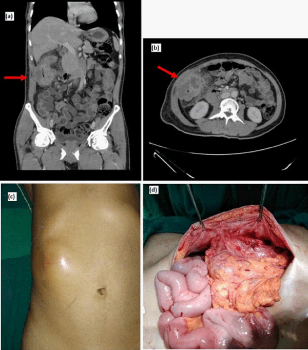
Patient was hence started on mesalazine, and advised to follow up. Patient was then lost to follow up for 4 months, when he presented again to the emergency department with fever with chills, right abdominal mass and had features of sepsis with tachycardia, elevated total counts and deranged liver & renal function tests. On examination, a well-defined firm, tender mass in the right hypochondrium and lumbar region extending up to right iliac fossa (Figure 1c).
Contrast enhanced computerized tomography of abdomen done showed heterogeneous wall thickening of the proximal part of the ascending colon with loss of mural stratification and causing luminal narrowing, measuring 5.56 x 4.28 x 4 cm with surrounding inflammatory changes with adjacent well defined collection 10.45 x 3.8 x 9.43 cms and few air pockets and infiltrating the right lateral abdominal wall (Figure 1a,b). Fine needle aspiration cytology from the abdominal swelling showed no evidence of malignant cells. Pus aspirated was sent for culture, grew Escherichia coli. As there was leukocytosis and left shift on complete blood picture, patient was given antibiotics according to culture sensitivity.
Patient was taken up for diagnostic laparoscopy and proceeded to exploratory laparotomy, abscess drained and an ileo-transverse colon anastomosis done. Intraoperatively omental deposits, and enlarged lymph nodes were noted (Figure 1c,d), FNAC done from these lesions were positive for malignancy, and histopathologcal examination was suggestive of metastatic mucin secreting adenocarcinoma.
Figure 1: a,b) CECT showing colonic growth with collection in abdominal wall. c) Subcutaneous abscess over right abdomen. d) Exploratory laparotomy picture showing growth infiltrating the abdominal wall.
Patient developed septicemia postoperatively. Patient had persistent hypotension and eventually expired, on the fourth postoperative day.
Colorectal cancer is a common and lethal disease, if not detected early and if treatment is delayed. The disease may progress without any symptoms early on, and also is notorious for its unusual presentations or presentation with discrete symptoms. It usually presents with rectal bleeding, abdominal pain, altered bowel habits, or anaemia and occult bleeding [6,7].
Unusual presentations include local invasion or a contained perforation leading to formation of a malignant fistula into adjacent organs (bladder or small bowel), pyrexia of unknown origin, intra-abdominal, retroperitoneal, abdominal wall or intrahepatic abscesses; Streptococcus bovis bacteremia and Clostridium septicum sepsis are associated with underlying colonic malignancies in approximately 10 to 25 percent of patients [8].
The conventional investigations like colonoscopy may be inadequate, as it cannot demonstrate abnormalities beyond the lumen of the colon. Repeat biopsies had shown benign lesions, non-specific pathology reports. Hence, a structured evaluation with computerized tomography (CT) of the abdomen is usually required. Exophytic growths, perforated cancers with local abscess formation and infiltration into adjacent structures, involvement of abdominal wall and its extent and lymph nodal involvement can be delineated on a CT. The CT scan in our case showed the presence of malignancy as well as an abscess, but didn’t show the relation between the two.
Occasionally a diagnostic laparoscopy or exploratory laparotomy maybe required to ascertain the diagnosis, and get a definitive pathological and microbiological sample for diagnosis, as done in our case.
Control of sepsis along with management of the primary malignancy is the usual line of treatment.
Our case highlights the importance of prompt diagnosis and early management, as the delay of 3 months caused extensive local spread of the disease and spreading sepsis which eventually led the patient to his death. Colonic Carcinoma should be considered in the differential diagnosis in the presentation of such an abscess, if accompanied by bowel symptoms. If not drained and resected, it can lead to complications like sepsis, which can endanger life.
- Three-years report of Population Based Cancer Registries 2006-2008 (Detailed Tabulations of Individual Registries Data). National Cancer Registry Programme (Indian Council of Medical Research), Bangalore November. 2010.
- World Cancer Research Fund and American Institute for Cancer Research Food, Nutrition, Physical Activity, and the Prevention of Cancer: A Global Perspective. Washington, DC: American Institute for Cancer Research. 2007.
- Merrill JG, Dockerty MB, Waugh JM. Carcinoma of the colon perforating onto the anterior abdominal wall. Surgery. 1950; 28: 662-671. PubMed: https://www.ncbi.nlm.nih.gov/pubmed/14782100
- Welch JP, Donaldson GA. Perforative carcinoma of the colon and rectum. Ann Surg. 1974; 180: 734-740. PubMed: https://www.ncbi.nlm.nih.gov/pubmed/4423043
- Andaz S, Heald RJ Abdominal wall abscess--an unusual primary presentation of a transverse colonic carcinoma. Postgrad Med J. 1993; 69: 826-828. PubMed: https://www.ncbi.nlm.nih.gov/pubmed/8290422
- Speights VO, Johnson MW, Stoltenberg PH, Rappaport ES, Helbert B, et al. Colorectal cancer: current trends in initial clinical manifestations. South Med J. 1991; 84: 575-578. PubMed: https://www.ncbi.nlm.nih.gov/pubmed/2035076
- Steinberg SM, Barkin JS, Kaplan RS, Stablein DM. Prognostic indicators of colon tumors. The Gastrointestinal Tumor Study Group experience. Cancer. 1986; 57: 1866-1870. PubMed: https://www.ncbi.nlm.nih.gov/pubmed/3485470
- Tsai HL, Hsieh JS, Yu FJ, Wu DC, Chen FM, et al. Perforated colonic cancer presenting as intra-abdominal abscess. Int J Colorectal Dis. 2007; 22: 15-19. PubMed: https://www.ncbi.nlm.nih.gov/pubmed/16625373
