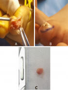
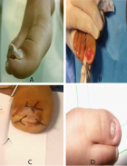
Figure 3
Subungual exostosis: Pediatric aspects
Mohamed Anouar Dendane, Achraf El Bakkaly*, Zakaria Alami Hassani and Abdelouahed Amrani
Published: 26 July, 2019 | Volume 3 - Issue 2 | Pages: 056-058
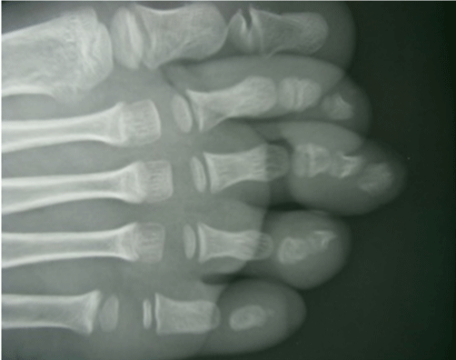
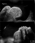
Figure 3:
X-ray image showing a bone outgrowth with lateral development and detached from the distal phalanx of the 3rd toe.
Read Full Article HTML DOI: 10.29328/journal.ascr.1001034 Cite this Article Read Full Article PDF
More Images
Similar Articles
-
Laparoscopic Adrenalectomy; A Short Summary with Review of LiteratureMushtaq Chalkoo*,Naseer Awan,Hilal Makhdoomi,Syed Shakeeb Arsalan ,Waseem Raja. Laparoscopic Adrenalectomy; A Short Summary with Review of Literature . . 2017 doi: 10.29328/journal.ascr.1001001; 1: 001-011
-
Bouveret Syndrome in an Elderly FemaleZvi H. Perry*, Udit Gibor,Shahar Atias,Solly Mizrahi,Alex Rosental, Boris Kirshtein. Bouveret Syndrome in an Elderly Female . . 2017 doi: 10.29328/journal.ascr.1001002; 1: 012-015
-
Intestinal obstruction complicated by large Morgagni herniaMartín Arnau B*,Medrano Caviedes R,Rofin Serra S, Caballero Mestres F,Trias Folch M. Intestinal obstruction complicated by large Morgagni hernia . . 2017 doi: 10.29328/journal.ascr.1001003; 1: 016-020
-
Clinical significance of Urinary Amylase in Acute PancreatitisMumtaz Din Wani,Mushtaq Chalkoo*,Zaheer Ahmed,Awhad Mueed Yousuf,Yassar Arafat,Syed Shakeeb Arsalan,Jaffar Hussain. Clinical significance of Urinary Amylase in Acute Pancreatitis . . 2017 doi: 10.29328/journal.ascr.1001004; 1: 021-031
-
Use of Orthodeoxia by pulse Oximetry in the detection of Hepatopulmonary SyndromeCesar Raul Aguilar Garcia*,Guadalupe Viridiana Ontiveros Guerra. Use of Orthodeoxia by pulse Oximetry in the detection of Hepatopulmonary Syndrome . . 2017 doi: 10.29328/journal.ascr.1001005; 1: 038-041
-
Surgery and new Pharmacological strategy in some atherosclerotic chronic and acute conditionsLuisetto M*,Nili-Ahmadabadi B,Ghulam Rasool Mashori. Surgery and new Pharmacological strategy in some atherosclerotic chronic and acute conditions . . 2017 doi: 10.29328/journal.ascr.1001006; 1: 042-048
-
The revolution of cardiac surgery evolution Running head: Cardiac surgery evolutionMarzia Cottini*. The revolution of cardiac surgery evolution Running head: Cardiac surgery evolution . . 2017 doi: 10.29328/journal.ascr.1001007; 1: 049-050
-
Dieulafoy’s Lesion related massive Intraoperative Gastrointestinal Bleeding during single Anastomosis Gastric Bypass necessitating total Gastrectomy: A Case ReportAshraf Imam,Khalayleh Harbi*,Miller Rafael, Khoury Deeb,Buyeviz Victor,Guy Pines,Sapojnikov Shimon. Dieulafoy’s Lesion related massive Intraoperative Gastrointestinal Bleeding during single Anastomosis Gastric Bypass necessitating total Gastrectomy: A Case Report . . 2017 doi: 10.29328/journal.ascr.1001008; 1: 051-055
-
Laparoscopic partial nephrectomy-does tumor profile influence the operative performance?Krishanu Das*, George P Abraham, Kishnamohan Ramaswai, Datson George P,Jisha J Abraham,Thomas Thachill, Oppukeril S Thampan. Laparoscopic partial nephrectomy-does tumor profile influence the operative performance? . . 2017 doi: 10.29328/journal.ascr.1001009; 1: 056-060
-
Comments for the Nuremberg Code 70 Years LaterJie Zhang,Chao-Jun Kong, Zhong Jia*. Comments for the Nuremberg Code 70 Years Later . . 2017 doi: 10.29328/journal.ascr.1001010; 1: 061-061
Recently Viewed
-
Advancing Forensic Approaches to Human Trafficking: The Role of Dental IdentificationAiswarya GR*. Advancing Forensic Approaches to Human Trafficking: The Role of Dental Identification. J Forensic Sci Res. 2025: doi: 10.29328/journal.jfsr.1001076; 9: 025-028
-
Scientific Analysis of Eucharistic Miracles: Importance of a Standardization in EvaluationKelly Kearse*,Frank Ligaj. Scientific Analysis of Eucharistic Miracles: Importance of a Standardization in Evaluation. J Forensic Sci Res. 2024: doi: 10.29328/journal.jfsr.1001068; 8: 078-088
-
Sinonasal Myxoma Extending into the Orbit in a 4-Year Old: A Case PresentationJulian A Purrinos*, Ramzi Younis. Sinonasal Myxoma Extending into the Orbit in a 4-Year Old: A Case Presentation. Arch Case Rep. 2024: doi: 10.29328/journal.acr.1001099; 8: 075-077
-
Toxicity and Phytochemical Analysis of Five Medicinal PlantsJohnson-Ajinwo Okiemute Rosa*, Nyodee, Dummene Godwin. Toxicity and Phytochemical Analysis of Five Medicinal Plants. Arch Pharm Pharma Sci. 2024: doi: 10.29328/journal.apps.1001054; 8: 029-040
-
Antibacterial Screening of Lippia origanoides Essential Oil on Gram-negative BacteriaRodrigo Marcelino Zacarias de Andrade, Bernardina de Paixão Santos, Roberson Matteus Fernandes Silva, Mateus Gonçalves Silva*, Igor de Sousa Oliveira, Sávio Benvindo Ferreira, Rafaelle Cavalcante Lira. Antibacterial Screening of Lippia origanoides Essential Oil on Gram-negative Bacteria. Arch Pharm Pharma Sci. 2024: doi: 10.29328/journal.apps.1001053; 8: 024-028.
Most Viewed
-
Evaluation of Biostimulants Based on Recovered Protein Hydrolysates from Animal By-products as Plant Growth EnhancersH Pérez-Aguilar*, M Lacruz-Asaro, F Arán-Ais. Evaluation of Biostimulants Based on Recovered Protein Hydrolysates from Animal By-products as Plant Growth Enhancers. J Plant Sci Phytopathol. 2023 doi: 10.29328/journal.jpsp.1001104; 7: 042-047
-
Sinonasal Myxoma Extending into the Orbit in a 4-Year Old: A Case PresentationJulian A Purrinos*, Ramzi Younis. Sinonasal Myxoma Extending into the Orbit in a 4-Year Old: A Case Presentation. Arch Case Rep. 2024 doi: 10.29328/journal.acr.1001099; 8: 075-077
-
Feasibility study of magnetic sensing for detecting single-neuron action potentialsDenis Tonini,Kai Wu,Renata Saha,Jian-Ping Wang*. Feasibility study of magnetic sensing for detecting single-neuron action potentials. Ann Biomed Sci Eng. 2022 doi: 10.29328/journal.abse.1001018; 6: 019-029
-
Pediatric Dysgerminoma: Unveiling a Rare Ovarian TumorFaten Limaiem*, Khalil Saffar, Ahmed Halouani. Pediatric Dysgerminoma: Unveiling a Rare Ovarian Tumor. Arch Case Rep. 2024 doi: 10.29328/journal.acr.1001087; 8: 010-013
-
Physical activity can change the physiological and psychological circumstances during COVID-19 pandemic: A narrative reviewKhashayar Maroufi*. Physical activity can change the physiological and psychological circumstances during COVID-19 pandemic: A narrative review. J Sports Med Ther. 2021 doi: 10.29328/journal.jsmt.1001051; 6: 001-007

HSPI: We're glad you're here. Please click "create a new Query" if you are a new visitor to our website and need further information from us.
If you are already a member of our network and need to keep track of any developments regarding a question you have already submitted, click "take me to my Query."


