Figure 1
A unique case of metastatic spinal epidural abscess associated with liver abscess following ascending cholangitis and Escherichia coli bacteremia
Nour Karra#, Samer Ganam#, Amitai Bickel, Maxim Bez, Ibrahim Abu Shakra, Doron Fischer and Eli Kakiashvili*
Published: 06 November, 2019 | Volume 3 - Issue 2 | Pages: 072-076

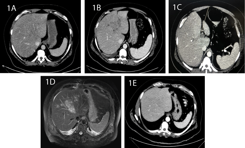
Figure 1:
CT images of the liver: 1A: Day 3. The shape and size are normal, with no pathological findings. 1B: Day 9. A hypodense irregular lesion is observed. 1C: Thrombus of the portal branch 1D: Abdominal MRI shows an extensive irregular process enhanced at T2, with multiple small cystic spaces at segments 2, 4 and 8. 1E: At day 24, the micro-abscesses are regressed following antibiotic treatment.
Read Full Article HTML DOI: 10.29328/journal.ascr.1001039 Cite this Article Read Full Article PDF
More Images
Similar Articles
-
Laparoscopic Adrenalectomy; A Short Summary with Review of LiteratureMushtaq Chalkoo*,Naseer Awan,Hilal Makhdoomi,Syed Shakeeb Arsalan ,Waseem Raja. Laparoscopic Adrenalectomy; A Short Summary with Review of Literature . . 2017 doi: 10.29328/journal.ascr.1001001; 1: 001-011
-
Bouveret Syndrome in an Elderly FemaleZvi H. Perry*, Udit Gibor,Shahar Atias,Solly Mizrahi,Alex Rosental, Boris Kirshtein. Bouveret Syndrome in an Elderly Female . . 2017 doi: 10.29328/journal.ascr.1001002; 1: 012-015
-
Intestinal obstruction complicated by large Morgagni herniaMartín Arnau B*,Medrano Caviedes R,Rofin Serra S, Caballero Mestres F,Trias Folch M. Intestinal obstruction complicated by large Morgagni hernia . . 2017 doi: 10.29328/journal.ascr.1001003; 1: 016-020
-
Clinical significance of Urinary Amylase in Acute PancreatitisMumtaz Din Wani,Mushtaq Chalkoo*,Zaheer Ahmed,Awhad Mueed Yousuf,Yassar Arafat,Syed Shakeeb Arsalan,Jaffar Hussain. Clinical significance of Urinary Amylase in Acute Pancreatitis . . 2017 doi: 10.29328/journal.ascr.1001004; 1: 021-031
-
Use of Orthodeoxia by pulse Oximetry in the detection of Hepatopulmonary SyndromeCesar Raul Aguilar Garcia*,Guadalupe Viridiana Ontiveros Guerra. Use of Orthodeoxia by pulse Oximetry in the detection of Hepatopulmonary Syndrome . . 2017 doi: 10.29328/journal.ascr.1001005; 1: 038-041
-
Surgery and new Pharmacological strategy in some atherosclerotic chronic and acute conditionsLuisetto M*,Nili-Ahmadabadi B,Ghulam Rasool Mashori. Surgery and new Pharmacological strategy in some atherosclerotic chronic and acute conditions . . 2017 doi: 10.29328/journal.ascr.1001006; 1: 042-048
-
The revolution of cardiac surgery evolution Running head: Cardiac surgery evolutionMarzia Cottini*. The revolution of cardiac surgery evolution Running head: Cardiac surgery evolution . . 2017 doi: 10.29328/journal.ascr.1001007; 1: 049-050
-
Dieulafoy’s Lesion related massive Intraoperative Gastrointestinal Bleeding during single Anastomosis Gastric Bypass necessitating total Gastrectomy: A Case ReportAshraf Imam,Khalayleh Harbi*,Miller Rafael, Khoury Deeb,Buyeviz Victor,Guy Pines,Sapojnikov Shimon. Dieulafoy’s Lesion related massive Intraoperative Gastrointestinal Bleeding during single Anastomosis Gastric Bypass necessitating total Gastrectomy: A Case Report . . 2017 doi: 10.29328/journal.ascr.1001008; 1: 051-055
-
Laparoscopic partial nephrectomy-does tumor profile influence the operative performance?Krishanu Das*, George P Abraham, Kishnamohan Ramaswai, Datson George P,Jisha J Abraham,Thomas Thachill, Oppukeril S Thampan. Laparoscopic partial nephrectomy-does tumor profile influence the operative performance? . . 2017 doi: 10.29328/journal.ascr.1001009; 1: 056-060
-
Comments for the Nuremberg Code 70 Years LaterJie Zhang,Chao-Jun Kong, Zhong Jia*. Comments for the Nuremberg Code 70 Years Later . . 2017 doi: 10.29328/journal.ascr.1001010; 1: 061-061
Recently Viewed
-
Agriculture High-Quality Development and NutritionZhongsheng Guo*. Agriculture High-Quality Development and Nutrition. Arch Food Nutr Sci. 2024: doi: 10.29328/journal.afns.1001060; 8: 038-040
-
A Low-cost High-throughput Targeted Sequencing for the Accurate Detection of Respiratory Tract PathogenChangyan Ju, Chengbosen Zhou, Zhezhi Deng, Jingwei Gao, Weizhao Jiang, Hanbing Zeng, Haiwei Huang, Yongxiang Duan, David X Deng*. A Low-cost High-throughput Targeted Sequencing for the Accurate Detection of Respiratory Tract Pathogen. Int J Clin Virol. 2024: doi: 10.29328/journal.ijcv.1001056; 8: 001-007
-
A Comparative Study of Metoprolol and Amlodipine on Mortality, Disability and Complication in Acute StrokeJayantee Kalita*,Dhiraj Kumar,Nagendra B Gutti,Sandeep K Gupta,Anadi Mishra,Vivek Singh. A Comparative Study of Metoprolol and Amlodipine on Mortality, Disability and Complication in Acute Stroke. J Neurosci Neurol Disord. 2025: doi: 10.29328/journal.jnnd.1001108; 9: 039-045
-
Development of qualitative GC MS method for simultaneous identification of PM-CCM a modified illicit drugs preparation and its modern-day application in drug-facilitated crimesBhagat Singh*,Satish R Nailkar,Chetansen A Bhadkambekar,Suneel Prajapati,Sukhminder Kaur. Development of qualitative GC MS method for simultaneous identification of PM-CCM a modified illicit drugs preparation and its modern-day application in drug-facilitated crimes. J Forensic Sci Res. 2023: doi: 10.29328/journal.jfsr.1001043; 7: 004-010
-
A Gateway to Metal Resistance: Bacterial Response to Heavy Metal Toxicity in the Biological EnvironmentLoai Aljerf*,Nuha AlMasri. A Gateway to Metal Resistance: Bacterial Response to Heavy Metal Toxicity in the Biological Environment. Ann Adv Chem. 2018: doi: 10.29328/journal.aac.1001012; 2: 032-044
Most Viewed
-
Evaluation of Biostimulants Based on Recovered Protein Hydrolysates from Animal By-products as Plant Growth EnhancersH Pérez-Aguilar*, M Lacruz-Asaro, F Arán-Ais. Evaluation of Biostimulants Based on Recovered Protein Hydrolysates from Animal By-products as Plant Growth Enhancers. J Plant Sci Phytopathol. 2023 doi: 10.29328/journal.jpsp.1001104; 7: 042-047
-
Sinonasal Myxoma Extending into the Orbit in a 4-Year Old: A Case PresentationJulian A Purrinos*, Ramzi Younis. Sinonasal Myxoma Extending into the Orbit in a 4-Year Old: A Case Presentation. Arch Case Rep. 2024 doi: 10.29328/journal.acr.1001099; 8: 075-077
-
Feasibility study of magnetic sensing for detecting single-neuron action potentialsDenis Tonini,Kai Wu,Renata Saha,Jian-Ping Wang*. Feasibility study of magnetic sensing for detecting single-neuron action potentials. Ann Biomed Sci Eng. 2022 doi: 10.29328/journal.abse.1001018; 6: 019-029
-
Pediatric Dysgerminoma: Unveiling a Rare Ovarian TumorFaten Limaiem*, Khalil Saffar, Ahmed Halouani. Pediatric Dysgerminoma: Unveiling a Rare Ovarian Tumor. Arch Case Rep. 2024 doi: 10.29328/journal.acr.1001087; 8: 010-013
-
Physical activity can change the physiological and psychological circumstances during COVID-19 pandemic: A narrative reviewKhashayar Maroufi*. Physical activity can change the physiological and psychological circumstances during COVID-19 pandemic: A narrative review. J Sports Med Ther. 2021 doi: 10.29328/journal.jsmt.1001051; 6: 001-007

HSPI: We're glad you're here. Please click "create a new Query" if you are a new visitor to our website and need further information from us.
If you are already a member of our network and need to keep track of any developments regarding a question you have already submitted, click "take me to my Query."


