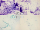
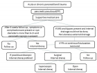
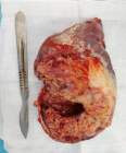
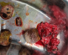
Figure 1
Cholecysto-colonic fistula after xanthogranulomatous cholecystitis: Surgeon’s nightmare
Karan Agarwal and Badareesh Lakshminarayana*
Published: 09 February, 2021 | Volume 5 - Issue 1 | Pages: 004-006
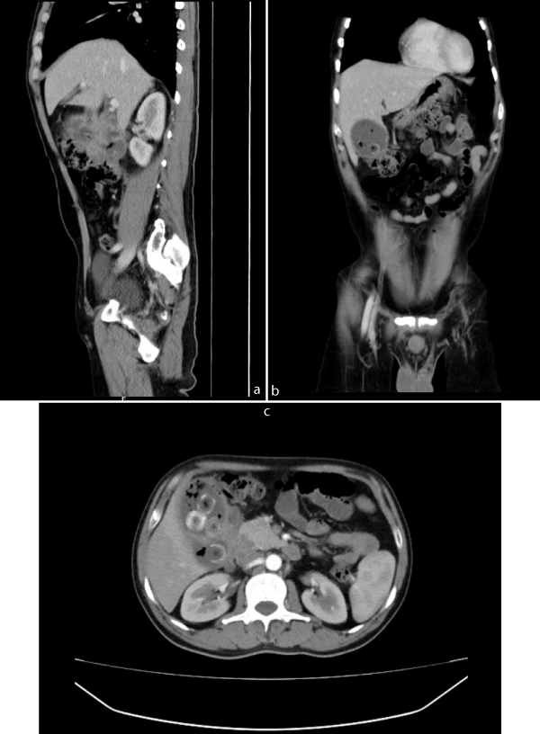
Figure 1:
1a-c: CECT abdomen and pelvis which showed distended Gall bladder with multiple calculi in the neck of gall bladder. Fat stranding and wall thickening of gall bladder is noted. Multiple air foci are noted within the lumen and along the wall of the gall bladder. Focal defect noted along the hepatic surface of neck of gall bladder with adjacent collection measuring 5.6 x 6.3 x 3.5 cm (AP x CC x TR) abutting the inferior surface of liver superolaterally, anterior pararenal space posteriorly, second part of duodenum medially, abutting and displacing hepatic flexure of colon inferiorly.
Read Full Article HTML DOI: 10.29328/journal.ascr.1001057 Cite this Article Read Full Article PDF
More Images
Similar Articles
-
Cholecysto-colonic fistula after xanthogranulomatous cholecystitis: Surgeon’s nightmareKaran Agarwal,Badareesh Lakshminarayana*. Cholecysto-colonic fistula after xanthogranulomatous cholecystitis: Surgeon’s nightmare. . 2021 doi: 10.29328/journal.ascr.1001057; 5: 004-006
Recently Viewed
-
Stages in COVID-19 vaccine development: The Nemesis, the Hubris and the ElpisVinod Nikhra*. Stages in COVID-19 vaccine development: The Nemesis, the Hubris and the Elpis. Int J Clin Virol. 2020: doi: 10.29328/journal.ijcv.1001028; 4: 126-135
-
Galenic hospital laboratory during COVID-19 emergency: A practical experience in an advanced countryLuisetto M*,Fiazza C,Ferraiuolo A,Ram Sahu. Galenic hospital laboratory during COVID-19 emergency: A practical experience in an advanced country. Int J Clin Virol. 2020: doi: 10.29328/journal.ijcv.1001027; 4: 118-125
-
COVID-19 and taking care and protection of patients with intellectual disabilities, need special care and equityMuhammad Tayyab*,Faheem Anwar,Jawad Khan,Ihteshamul Haq. COVID-19 and taking care and protection of patients with intellectual disabilities, need special care and equity. Int J Clin Virol. 2020: doi: 10.29328/journal.ijcv.1001026; 4: 116-117
-
COVID-19 pandemic, recurrent outbreaks and prospects for assimilation of hCoV-19 into the human genomeVinod Nikhra*. COVID-19 pandemic, recurrent outbreaks and prospects for assimilation of hCoV-19 into the human genome. Int J Clin Virol. 2020: doi: 10.29328/journal.ijcv.1001025; 4: 111-115
-
The expected second wave of COVID-19Madiha Asghar*,Misbahud Din. The expected second wave of COVID-19. Int J Clin Virol. 2020: doi: 10.29328/journal.ijcv.1001024; 4: 109-110
Most Viewed
-
Evaluation of Biostimulants Based on Recovered Protein Hydrolysates from Animal By-products as Plant Growth EnhancersH Pérez-Aguilar*, M Lacruz-Asaro, F Arán-Ais. Evaluation of Biostimulants Based on Recovered Protein Hydrolysates from Animal By-products as Plant Growth Enhancers. J Plant Sci Phytopathol. 2023 doi: 10.29328/journal.jpsp.1001104; 7: 042-047
-
Sinonasal Myxoma Extending into the Orbit in a 4-Year Old: A Case PresentationJulian A Purrinos*, Ramzi Younis. Sinonasal Myxoma Extending into the Orbit in a 4-Year Old: A Case Presentation. Arch Case Rep. 2024 doi: 10.29328/journal.acr.1001099; 8: 075-077
-
Feasibility study of magnetic sensing for detecting single-neuron action potentialsDenis Tonini,Kai Wu,Renata Saha,Jian-Ping Wang*. Feasibility study of magnetic sensing for detecting single-neuron action potentials. Ann Biomed Sci Eng. 2022 doi: 10.29328/journal.abse.1001018; 6: 019-029
-
Pediatric Dysgerminoma: Unveiling a Rare Ovarian TumorFaten Limaiem*, Khalil Saffar, Ahmed Halouani. Pediatric Dysgerminoma: Unveiling a Rare Ovarian Tumor. Arch Case Rep. 2024 doi: 10.29328/journal.acr.1001087; 8: 010-013
-
Physical activity can change the physiological and psychological circumstances during COVID-19 pandemic: A narrative reviewKhashayar Maroufi*. Physical activity can change the physiological and psychological circumstances during COVID-19 pandemic: A narrative review. J Sports Med Ther. 2021 doi: 10.29328/journal.jsmt.1001051; 6: 001-007

HSPI: We're glad you're here. Please click "create a new Query" if you are a new visitor to our website and need further information from us.
If you are already a member of our network and need to keep track of any developments regarding a question you have already submitted, click "take me to my Query."


